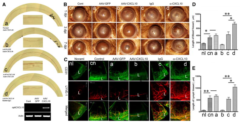
Fig. 1.
Epithelial levels of CXCL10 control C. albicans induced angiogenesis in the cornea. A. AAV-9 mediated epithelial expression of CXCL10 in the cornea. AAV9-GFP or AAV9CXCL10 were injected subconjunctivally and at day 14 corneas were collected and the expression of CXCL10 was assessed using immunohistochemistry and RT-PCR. B. To assess the role of exogenously expressed or endogenously induced CXCL10, AAV9-GFP or AAV9CXCL10, CXCL10 neutralizing antibody, or nonspecific IgG as the control, was injected subconjunctuvally. The treated corners, 14 days post AAV9 infection or 6 h post antibody injection, were inoculated with C. albicans and photographed at the indicated days p.i. with slid lamp. C. The blood and lymph vessels were visualized by whole mount staining of the corneas with CD31 for blood and LYVE1 for lymph vessels. L: Limbus. To quantify neovascularization in infected corneas, the lengths of blood (D) or lymph vessels (E) from the edge of the limbal region to the tip of vessels was measured by Image J. The results are representative of two independent experiments (N = 3 each) and indicated p values were generated using unpaired Student’s t test. **p < 0.01, *p < 0.05

