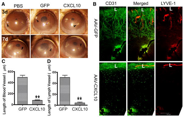
Fig. 4.
CXCL10 blocks suture-induced angiogenesis in the cornea. B6 mouse corneas were infected with AAV9-GFP or AAV9CXCL10. At day 14 post AAV9 infection, three non-penetrating sutures were placed. A. At 3 (3d) and 7 (7d) days post suture, corneas were photographed. Arrowheads, suture sites. B. At 7 days post suture, the corneas were excised, and subjected to whole mount staining with CD31 for blood (green) and LYVE1 for lymph (red) vessels, respectively. L: limbus. C. Quantitation of the lengths of blood vessels from the edge of the limbal region to the tips of vessels in the infected corneas. D. Quantitation of the lengths of lymph vessels from the edge of the limbal region to the tips of vessels in the corneas. The results are representative of two independent experiments (N = 3 each) and indicated p values were generated using unpaired Student’s t test. **p < 0.01, *p < 0.05. (Color figure online)

