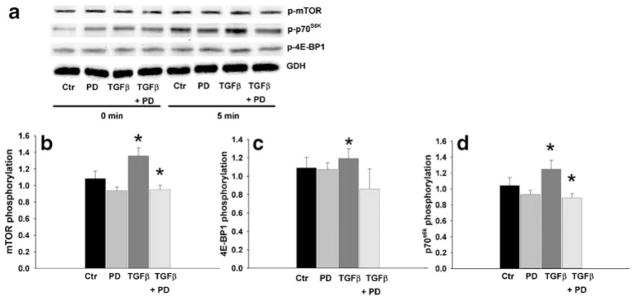
Fig. 6.
TGFβ stimulated mTOR, p70S6K, and 4E-BP1 phosphorylation in cells treated for 5 min is abolished by pre-treatment with the MEK inhibitor PD98059 (10 μM) for 1 h. a Typical Western blot probed for phosphorylated mTOR, phosphorylated p70S6K, phosphorylated 4E-BP1, and GAPDH for the indicated times. b–d Densitometric results as the ratio of phosporylated protein normalized to GAPDH (n≥4, *P<0.05). Control phosphorylation (Ctr) at time point 0 was defined at 1.0

