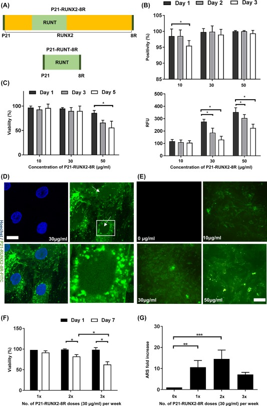
Figure 1.

Efficient delivery and dosing of P21‐RUNX2‐8R to initiate osteogenesis in human mesenchymal stem cells (hMSCs). (A): Design of the osteogenic constructs. P21‐RUNX2‐8R is a RUNX2 transcription factor with an N‐terminal fusion of P21 and a C‐terminal fusion of 8R. P21‐RUNT‐8R contains only the DNA binding domain, RUNT, sandwiched between P21 and 8R. (B): To assess the delivery of the fusion peptide, the proteins were labeled with FITC and delivered at different concentrations overnight. Flow cytometry analysis of percentage positivity and relative fluorescence unit of hMSCs treated with different concentrations of P21‐RUNX2‐8R‐FITC (10, 30, 50 μg/ml) overnight. Statistical significance was determined using the Holm‐Sidak method, α = 0.05;*, p < .05. (C): Viability of hMSCs measured using trypan blue at day 1 and 7 after overnight treatment with 30 μg/ml of P21‐RUNX2‐8R. Thirty μg/ml of P21‐RUNX2‐8R was used as the optimal concentration on further experiments. Statistical significance determined using the Holm‐Sidak method, with α = .05; *, p < .05. (D): Confocal images of hMSCs treated with 30 μg/ml P21‐RUNX2‐8R‐FITC overnight and counter stained with Hoechst (nuclei stain) at ×40 magnification. Significant amounts of P21‐RUNX2‐8R‐FITC are colocalized with the Hoechst stained cell nucleus. Most P21‐RUNX2‐8R‐FITC is localized in small endosomal vesicles present in perinuclear region in the cytoplasm (hatched area). Scale bar is 10 μm. (E): Fluorescence microscopy images of hMSCs treated with 10, 30, and 50 μg/ml P21‐RUNX2‐8R‐FITC overnight. As the concentration increases, fluorescence intensity of P21‐RUNX2‐8R‐FITC inside hMSCs increases. Scale bar is 50 μm. (F): For the first week, hMSCs were treated overnight with P21‐RUNX2‐8R (30 μg/ml) every other night (3×), every 3 days (2×), or treated only once (1×). Viability of hMSCs measured using trypan blue at day 1 and 7 after overnight treatment with 30 μg/ml of P21‐RUNX2‐8R once, twice and thrice per week. Statistical significance determined using the Holm‐Sidak method, with α = 0.05; *, p < .05. (G): Treated hMSCs were cultured in osteo‐permissive medium for 3 weeks, stained with alizarin red S (ARS) for matrix mineralization and quantified using a microplate reader. Thirty μg/ml of P21‐RUNX2‐8R treated twice during the first week was identified as the optimal dose, and the same conditions were used in the subsequent assays. Statistical significance was determined using one way ANOVA, with α = 0.05;*, p < .05; **, p < .005; ***, p < .001. Error bars indicate standard deviation (SD). hMSCs from two different donors were used for this study.
