Figure 3.

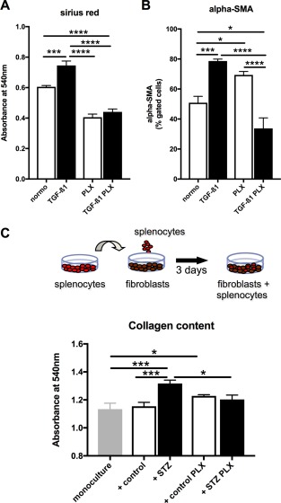
PLX exert antifibrotic effects in vitro. PLX conditioned medium reduces TGF‐β1‐induced collagen deposition and α‐SMA expression in cardiac fibroblasts. Bar graphs represent the mean ± SEM of (A) the absorbance at 540 nm detected after Sirus Red staining, depicting collagen deposition, and (B) α‐SMA expression of cardiac fibroblasts cultured in PLX medium, PLX medium + TGF‐β1, PLX conditioned (cond.) medium, or PLX cond. medium + TGF‐β1 as indicated, with open bars representing without TGF‐β1 and black bars with TGF‐β1 conditions, and n = 6/group. Splenocytes derived from STZ PLX mice induce lower collagen deposition in murine fibroblasts upon coculture compared with splenocytes from STZ mice. (C): Schematic representation of the experimental set‐up. (D): Bar graph represents the mean ± SEM of the absorbance at 540 nm detected after Sirus Red staining of fibroblasts cultured in monoculture or cocultured with splenocytes from control, STZ, control PLX, or STZ PLX mice as indicated, with n = 5–6/group. For all data: *, p < .05; **, p < .01; ***, p < .001; ****, p < .0001. Abbrevaitions: PLX, PLacenta‐eXpanded cell; STZ, streptozotocin; TGF‐ß1, transforming growth factor‐β1.
