
Figure 3.
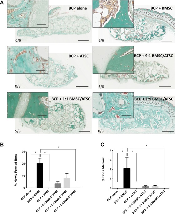
Ectopic bone formation by BCP biomaterial with MSC in the subcutis of nude mice after 8 weeks. (A): Masson trichrome staining on sections through implants with BCP (gray) and MSC from three bone marrow donors (BMSC) and four adipose tissue donors (ATSC), or a mixture of both. Newly formed bone is evidenced in green. Scale bars: 1 mm for images (40 μm for image inserts). The bone incidence score on each group represents the number of implants with newly formed ectopic bone over the total number of implants in that group. (B): Histomorphometry quantification of bone and (C) of mature bone marrow was performed on four sections of every implant. * indicates statistical differences between groups (p < .05). Data are presented as mean ± SE of the mean. Abbreviations: ATSC, adipose tissue‐derived stem cells; BCP, biphasic calcium phosphate; BMSC, bone marrow‐derived stem cells.