
Figure 6.
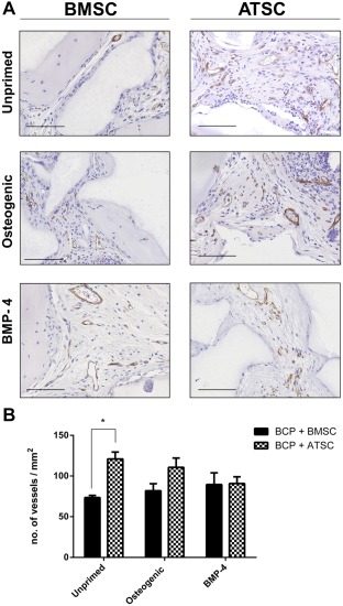
Neovascularization within implants of BCP with MSC in the subcutis of nude mice after 8 weeks. (A): CD146 immunostaining for pericytes on sections through implants with BCP biomaterial (gray) and MSC from three bone marrow donors (BMSC) and four adipose tissue donors (ATSC) which were either unprimed or underwent priming osteogenic supplements (250 μM ascorbic acid, 10 mM β‐glycerolphosphate, and 10 nM dexamethasone) or 50 ng/ml rh BMP‐4 prior to implantation. Blood vessels are stained in brown. Scale bars represent 100 μm. (B): Histomorphometry quantification of the number of blood vessels/mm2 with * indicating statistical differences between groups (p < .05). Data are presented as mean ± SE of the mean. Abbreviations: ATSC, adipose tissue‐derived stem cells; BCP, biphasic calcium phosphate; BMP‐4, bone morphogenetic protein 4; BMSC, bone marrow‐derived stem cells.