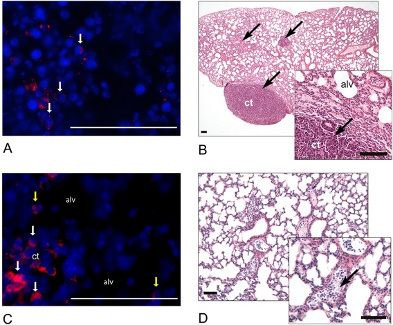
Figure 2.

Histological analysis of tissues following C91–98 cell injections. (A): Presence of GFP‐immunoreactivity (red, arrows) in the lumps at the site of injection, nuclei—blue. (B): A representative lung section demonstrating areas of ct on D28 (Mayer's hematoxylin and eosin). Ct areas are indicated with arrows. (C): A representative lung section demonstrating the presence of GFP immunoreactivity within the ct (white arrows) and in the alveolar region (alv, yellow arrows) on D28: GFP (red), Hoechst (blue). (D): Nucleated cells in lung blood vessel lumena (Mayer's hematoxylin and eosin). The presence of nucleated cells is marked with an arrow within the enlarged inset. Scale = 100 µm. Abbreviations: alv, alveolus; ct, condensed tissue; GFP, green fluorescent protein.
