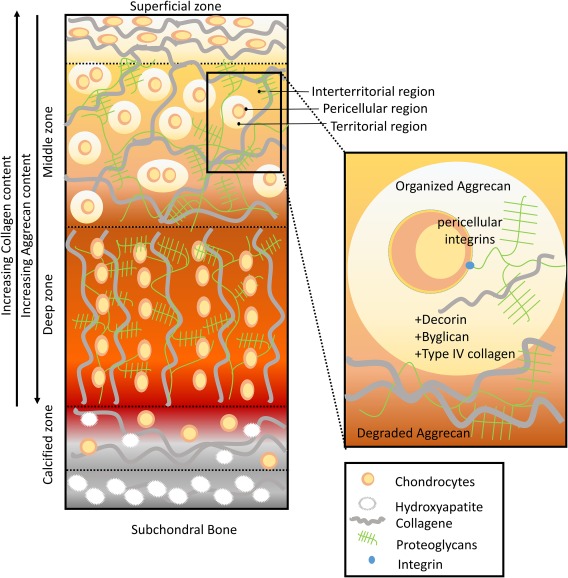
Figure 2.

Human articular cartilage. Articular cartilage is divided into four different zones: superficial, middle, deep, and calcified zone. The four zones differ in their collagen, cell orientation, and proteoglycan density. In the superficial zone, collagen fibers are thin and organize themselves parallel to the plane of the articular surface. In the middle zone, the collagen fibers appear more randomly oriented. In the deep zone collagen fibers become thicker and align orthogonally to the superficial zone. At the same time aggrecan density increases from the superficial zone toward the deep zone. Also chondrocyte morphology and density changes depending on the zone. In the superficial zone, chondrocytes are disc‐shaped, aligned parallel to the articular surface as well as the collagen fibers. The middle zone is characterized by randomly oriented spherical cells that could be either isolated or in small clusters. In the deep zone chondrocytes are ellipsoid and aligned in columns. Cartilage's extracellular matrix (ECM) is differentially organized around the chondrocytes in pericellular, territorial, and interterritorial region. These different ECM organizations serve to protect the cells and transmit mechanical signals. The pericellular zone is full of proteoglycans (byglicans, aggrecans). The territorial zone also contains thin collagen fibrils. Instead, the interterritorial matrix constitutes 90% of cartilage volume and contains the largest collagen fibrils.
