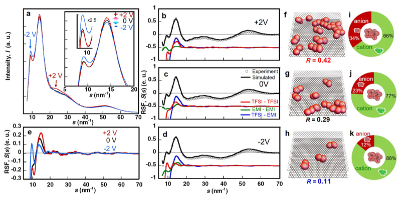
Figure 6. The effect of electrode potential on the structural ordering of ionic liquid inside ultra-narrow pores.
(a) Change in In-situ X-ray scattering profile of EMI-TFSI in the monolayer confinement of carbon nanopores of 0.7 nm under constant potentials of 0 V (black) and ± 2 V (+: red and -: blue). Here, we express application of the electric potentials of 2 V for positive and negative electrodes as +2 V and -2 V, respectively. (b, c, d) Experimental X-ray Reduced Structure Functions (RSFs) of EMI-TFSI in the 0.7-nm pores under +2 V (b), 0 V (c) and -2 V (d) with open circles. The HRMC-simulated RSFs are plotted with black solid lines. The simulated RSFs for the TFSI-TFSI correlation (red), EMI-EMI correlation (green) and TFSI-EMI correlation (blue) are given as solid lines. (e) Single plots of RSFs of TFSI-TFSI correlation in the 0.7-nm pores under +2 V (red), 0 V (black) and -2 V (blue). (f, g, h) Snapshots of co-ion pairs of anions of EMI-TFSI in the 0.7 nm-pore under +2 V (f), 0 V (g) and -2 V (h). R is the ratio of paired anion number to total anion number. TFSI anions are shown as red ellipsoids. (i, j, k) Population in the first coordination shell around a TFSI anion in the 0.7-nm pore under +2 V (i), 0 V (j) and -2 V (k).

