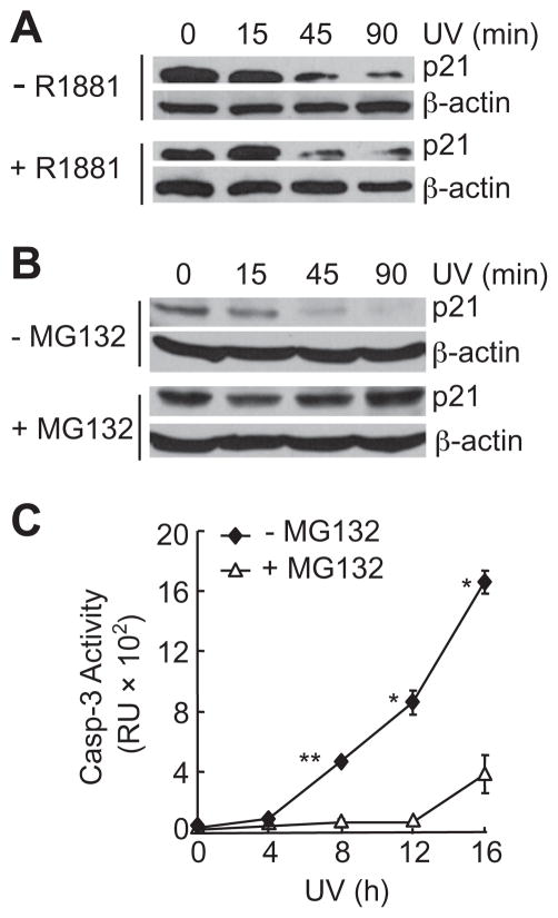
Fig. 1.
UV-induced degradation of p21 protein in AR-positive 104-R cells. (A) 104-R cells were treated with or without a synthetic androgen, R1881 (2 nM) for 2 h followed by UV irradiation (100 J/m2). The p21 protein levels were analyzed by immunobloting with anti-p21 antibody. (B and C) 104-R cells were treated with or without proteasome inhibitor MG-132 (10 μM) for 2 h. The p21 protein levels were analyzed by immunoblotting (B), as described in (A). Caspase activity was measured using fluorogenic substrate Ac-DEVD-AFC (C). *p < 0.05, **p < 0.01.

