Fig. 2.
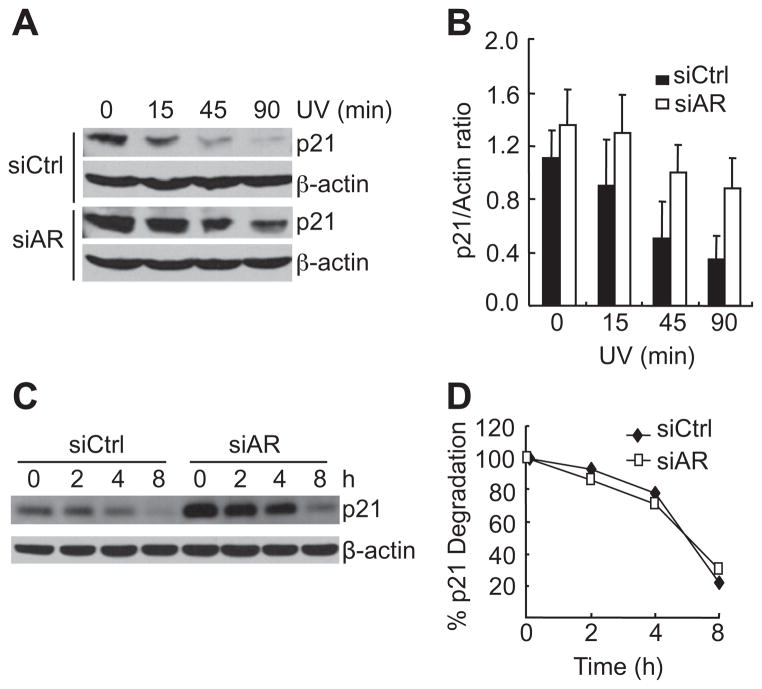
AR controls basal p21 expression, but not its degradation rate. (A) 104-R cells stably transfected with either control siRNA (siCtrl) or AR siRNA (siAR) [14]. Cells were irradiated by UV (100 J/m2) for various periods of time, as indicated. p21 protein levels were analyzed by immunoblotting with anti-p21 antibody. (B) Quantitation of p21 protein levels using image J software. The data are from three independent experiments (C) 104-R cells were treated with MG-132 (10 μM) for 12 h, completely washed, and then treated with CHX (50 μg/ml) for various periods of time, as indicated. The p21 protein levels were analyzed by immunoblotting with anti-p21 antibody. (D) Quantitation of p21 protein levels using image J software.

