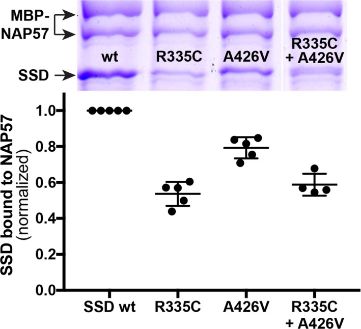
Figure 2.

Pulldowns of recombinant wild type (wt) and mutant SSD with full‐length NAP57 fused to MBP using amylose beads. (Above) Representative coomassie blue stained denaturing polyacrylamide gel with the bands labeled on the left. Note the MBP‐NAP57 migrates as a doublet. (Below) Quantification of the SSD bands relative to MBP‐NAP57. Between 4 and 5 independent experiments were normalized to wild type SSD. In the last lane, the two variant proteins were mixed 1:1.
