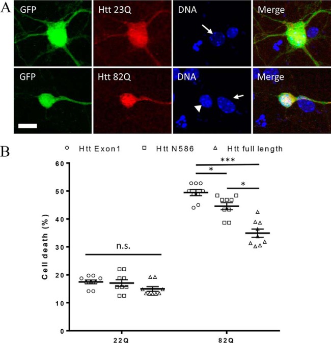
Figure 1.
Neuronal toxicity of expanded full-length Htt. A, example images of primary neurons transfected with full-length Htt containing either 23Q or 82Q and co-transfected with GFP. Transfected Htt was detected by immunocytochemistry using mAb 2166, and DNA is stained with Hoechst. Condensed nuclei (arrowhead) are brighter and smaller than healthy nuclei (arrow). B, quantification of cell death by nuclear condensation assay. Primary cortical neurons were transfected at DIV5 with indicated plasmid and with GFP. The cells were fixed after 48 h, and the nuclei were stained with Hoechst. Automated picture acquisition was performed on Zeiss Axiovert 200 microscope, and automated quantification of the nuclear intensity of transfected cells was performed using Volocity. *, p ≤ 0.05; ***, p ≤ 0.001 (n = 9 independent neuronal preparations).

