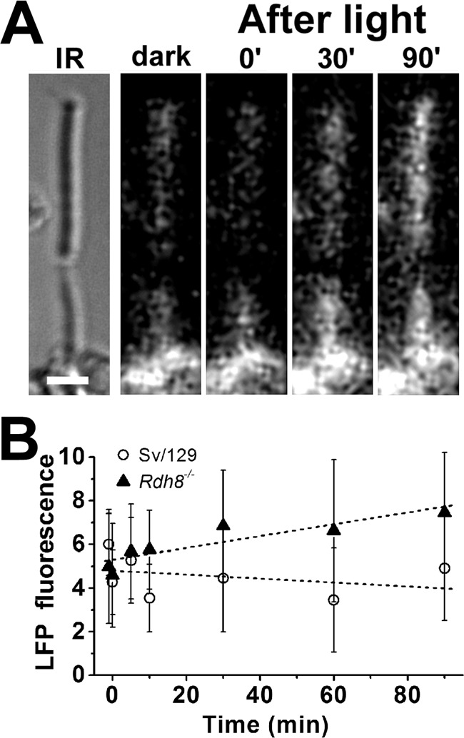
Figure 9.

Formation of LFP in metabolically intact rod photoreceptors of Rdh8−/− mice. A, IR, infrared image of a rod photoreceptor cell isolated from an Rdh8−/− mouse; bar, 5 μm. Bleaching was carried out between t = −1 min and t = 0. Fluorescence images were acquired with 490 nm excitation and >515 nm emission. B, outer segment LFP fluorescence (excitation, 490 nm; emission, >515 nm) after bleaching in rod photoreceptors isolated from Rdh8−/− mice (▴, n = 12). Data from wild-type mice (○, n = 11) are re-plotted from Ref. 35 for comparison. Error bars represent standard deviations.
