Figure 4.
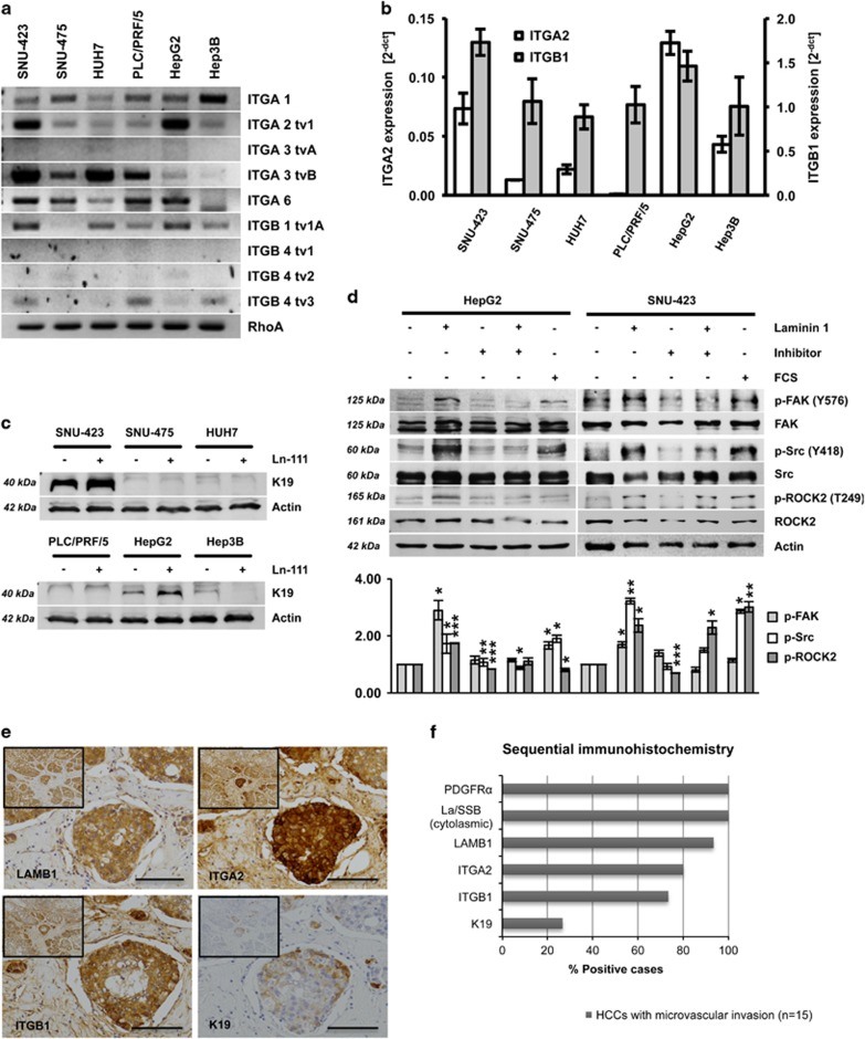
Ln-111 activates ITG signaling and downstream ROCK2. (a) HCC cell lines were analyzed for ITG receptor expression by PCR or (b) qPCR. (c) K19 levels of HCC cell lines cultured on plastic or Ln-111-coated dishes were detected by western blot analysis. (d) HepG2 and SNU-423 cells were cultured on plastic or Ln-111-coated dishes in the presence or absence of an inhibitory antibody specific for Ln-111. The medium was replaced with serum-free medium after 12 h. Lysates were taken after 24 h and analyzed by western blotting. For the fetal calf serum (FCS)-positive control, 10% FCS were added to the media 30 min before cell lysis. Ln-111 coating induced the phosphorylation of ITG-specific downstream proteins focal adhesion kinase (FAK), Src and ROCK2. The diagram in the lower panel shows relative values of pFAK/FAK, pSrc/Src and pROCK2/ROCK2 of the quantified western blot (n=3). Signal intensities of untreated cells were set to a value of 1 for normalization. (e) Representative HCC sample showing strong positivity for LAMB1, ITGA2 and ITGB1, and focal positivity for K19 in invading tumor cells located in a blood vessel (scale bars 100 μm). (f) Sequential stainings for PDGFRα, cytosolic La/SSB, LAMB1, ITGA2, ITGB1 and K19 were performed on 15 HCC samples with microvascular invasion and scored for immunopositivity. All data are presented as means±s.d. and analyzed using the Student’s t-test (*P<0.05, **P<0.01, ***P<0.005).

