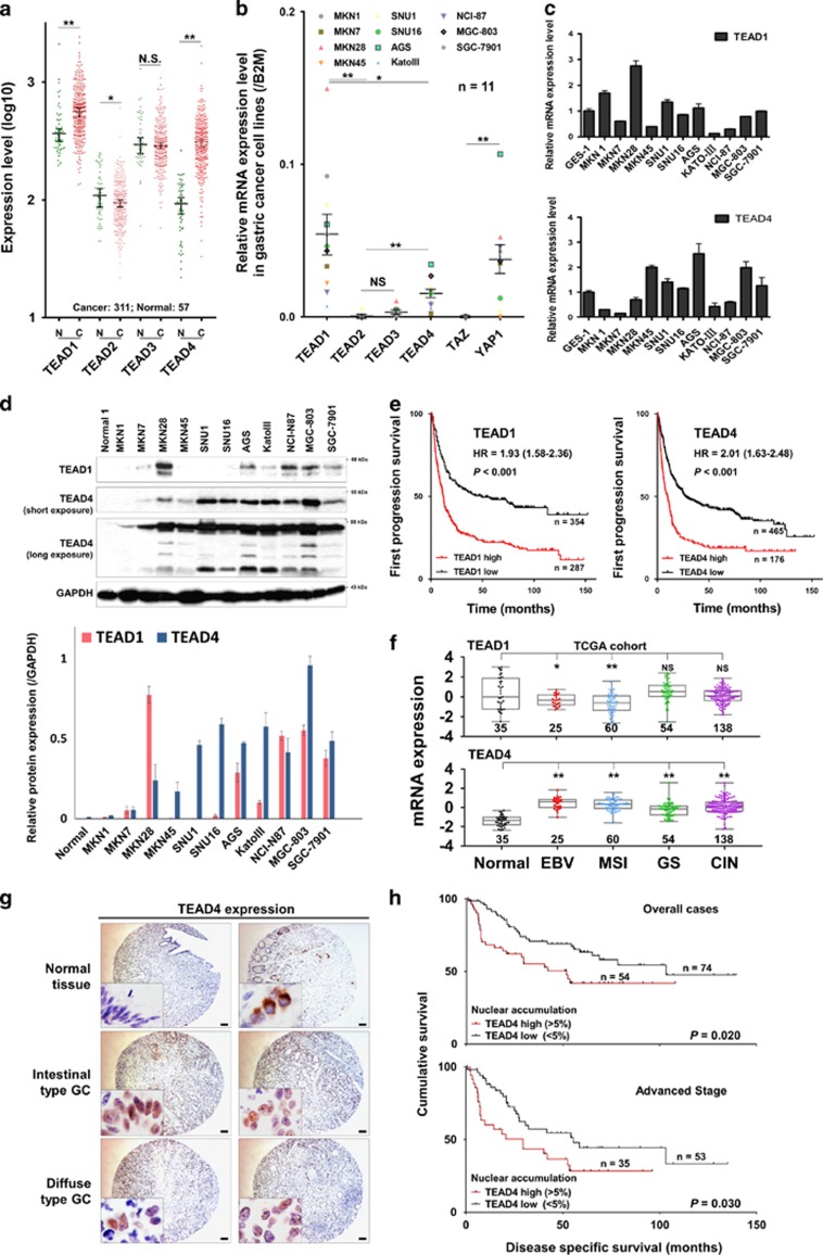
Figure 1.
TEAD1 and TEAD4 are upregulated and predict poor prognosis in GC. (a) TEAD1 and TEAD4 displayed high expression in 311 GC samples compared with normal gastric tissues (http://medical-genome.kribb.re.kr/GENT/; *P<0.05; **P<0.001; NS, not significant; unpaired t-test). (b) TEAD1 and TEAD4 were upregulated in GC cell lines compared with TEAD2/3; meanwhile, expression of YAP1 is significantly higher than that of TAZ (**P<0.001; unpaired t-test). (c) mRNA expression of TEAD1 and TEAD4 in 11 GC cell lines compared with immortalized gastric epithelium cell line GES-1. (d) High protein level of TEAD1 and TEAD4 was detected in most of the GC cell lines (upper panel). And the corresponding quantification by densitometry was shown at nether panel. (e) Overexpressed TEAD1 and TEAD4 were associated with poor progression-free survival in primary GCs (P<0.001) from Kaplan–Meier plotter (NCBI/GEO/GSE14210, GSE15459, GSE22377, GSE29272, GSE51105 and GSE62254). (f) Distribution of TEAD1 and TEAD4 mRNA expression in four molecular subtypes of GC (*P<0.05; **P<0.001; TCGA cohort; unpaired t-test). (g) Immunohistochemistry images of TEAD4 in GC tissue microarray (Scale bars are 200 μm). TEAD4 exhibited negative or cytoplasmic expression in normal epithelium cells, but it was localized in the nucleus of the cancer cells. (h) High TEAD4 nuclear accumulation was associated with poor disease-specific survival (overall cases, P=0.020; advanced-stage cases, P=0.030; log-rank (Mantel–Cox) test).

