Figure 2.
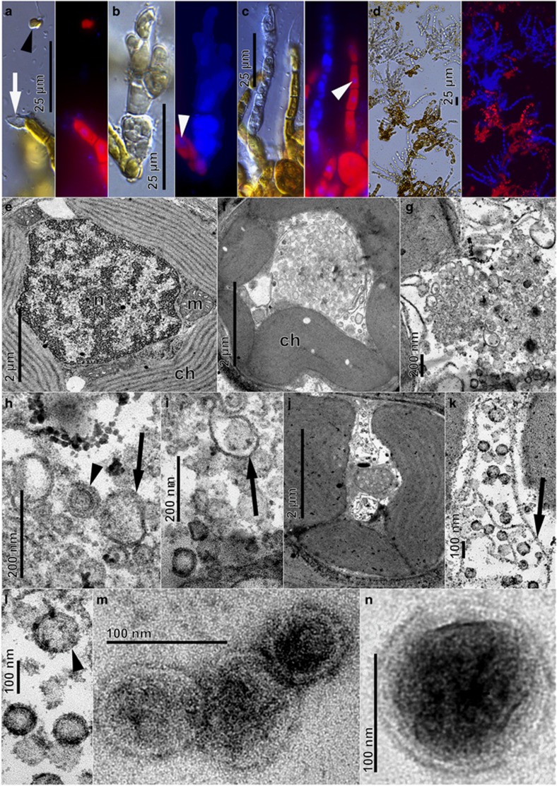
Light and epifluorescence (a–d, DAPI stained) and transmission electron (e–n) micrographs of Laminaria digitata gametophyte strain LdigPH10-30 m. (a) Spermatozoid (arrowhead) released from antheridium (white arrow), (b,c). Deformed opaque structures with high DAPI blue fluorescence in contrast to normal nuclei (white arrowheads). (d) High prevalence of DAPI-fluorescent filaments. (e) Cross-section of healthy vegetative cell showing chloroplast (ch), nucleus (n), and mitochondria (m). (f–l) VLP formation in vegetative gametophyte cells. Chloroplasts detached from cell periphery, loss of internal structure, appearance of tubular structures (arrows) and various stages of VLP assembly (arrowheads). (m,n) VLPs isolated from extracellular medium and visualised by negative staining.

