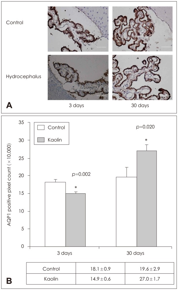
FIGURE 3. Aquaporin (AQP) 1 immunolabeling in the choroid plexus. (A) Immunohistochemical staining and (B) quantificative measurement by positive pixel count of AQP 1 of brain parenchyma revealed expression to be higher than in the control at 3 days and 30 days (n=3, *p<0.05).

