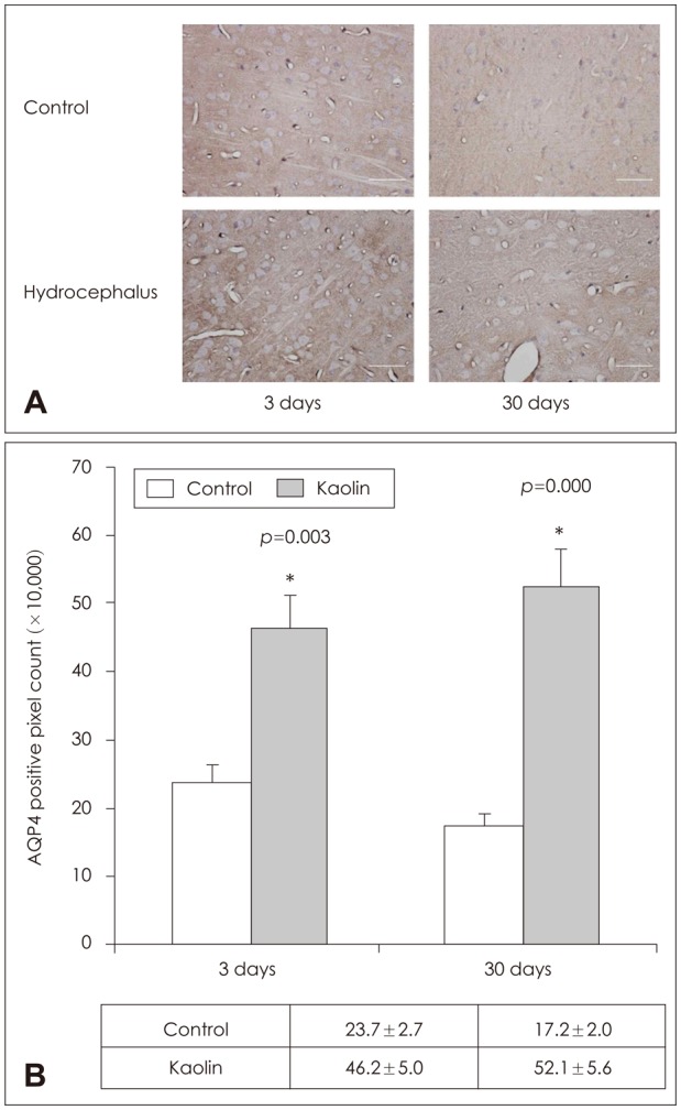
FIGURE 5. Aquaporin (AQP) 4 immunolabeling in the parenchyme. (A) Immunohistochemical staining and (B) quantificative measurement by positive pixel count of AQP4 of the brain parenchyma revealed expression to be higher than in the control at day 3 and day 30 (n=3, *p<0.05).

