. 2017 Oct 31;13(2):76–84. doi: 10.13004/kjnt.2017.13.2.76

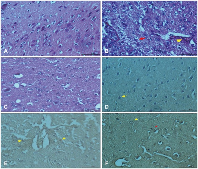
FIGURE 1. (A) Normal appearance of regular cells and vascular structures in brain cortex, hematoxylin and eosin (H & E) staining bar 50 µm (control group). (B) Dilation in blood vessels and hemorrhage (arrowhead), degeneration in some neuron (arrow) H & E staining bar 50 µm (trauma group). (C) Reduction in vascular dilatation, regular structure of the nucleus and cytoplasm in neurons and glial cells, H & E staining bar 50 µm (trauma + Ganoderma group). (D) Expression of phosphorylated p38 mitogen activated protein kinase (MAPK) in neuron nucleus (arrow), phosphorylated p38 immunohistochemistry (IHC) staining bar 50 µm (control group). (E) Positive expression of phosphorylated p38 MAPK in nucleus of neuron and glia cells (arrows), phosphorylated p38 IHC staining bar 50 µm (trauma group). (F) Increased p38 expression in neuronal membranes (yellow arrow) and nucleus of glial cells phosphorylated p38 (red arrow) IHC staining bar 50 µm (trauma+Ganoderma group).

FIGURE 1