. 2017 Oct 31;13(2):76–84. doi: 10.13004/kjnt.2017.13.2.76

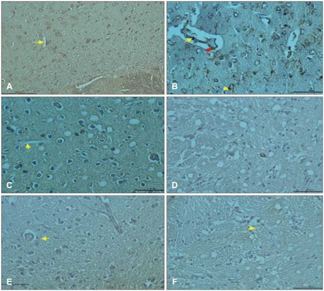
FIGURE 2. (A) Vascular endothelial growth factor (VEGF) expression of vascular endothelial cells in the cortex (arrow) VEGF immunohistochemistry staining bar 50 µm (control group). (B) Positive VEGF expression in endothelial cells and inflammatory cells (arrows) VEGF immunohistochemistry staining bar 50 µm (trauma group). (C) Increased VEGF expression in endothelial cells (arrow), VEGF immunohistochemistry staining bar 50 µm (trauma+Ganoderma group). (D) Positive cluster of differentiation 68 (CD68) expression in glial cells surrounding of the blood vessels, CD68 immunohistochemistry staining bar 50 µm (control group). (E) Weak CD68 expression in glial cells (arrow) CD68 immunohistochemistry staining bar 50 µm (trauma group). (F) Positive CD68 expression in glia cells around blood vessels (CD68 immunohistochemistry staining bar 50 µm) (trauma+Ganoderma group).

FIGURE 2