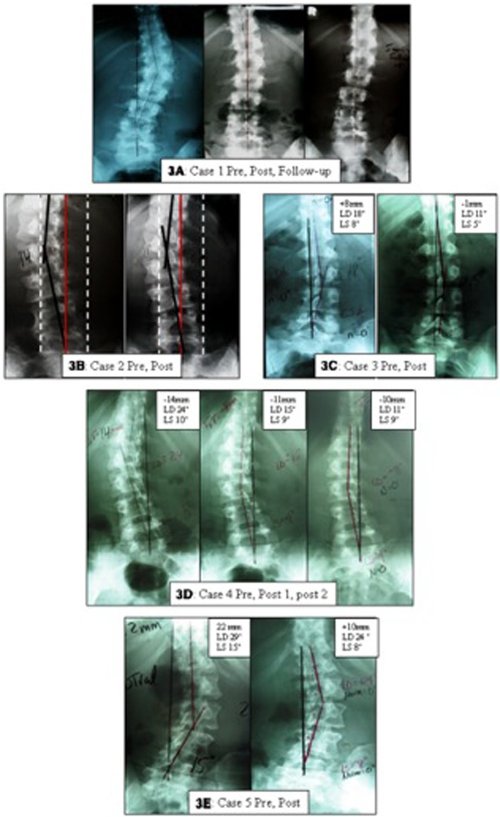
Fig. 3.
Initial and follow-up lumbo-pelvic radiographs of the five cases with scoliosis. In A, a 17 year old female’s initial, 72 visit post analysis, and 5-month follow-up AP lumbo-pelvic x-rays are shown. In B, a 35 year old female’s initial and 72 visit post analysis AP lumbo-pelvic x-rays are shown. In C, a 19 year old female’s initial and 18 visit post AP lumbo-pelvic x-rays are shown. In D, a 41 year old female’s initial, 36 visit, and 84 visit post analysis AP lumbo-pelvic x-rays are shown. In E, a 45 year old female’s initial and 36 visit post analysis AP lumbo-pelvic x-rays are shown.

