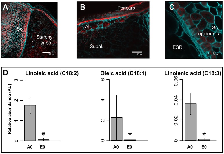
Figure 2.
Precise localization of oil bodies and compartmentation of fatty acids between rice seed tissues. (A–C) Lipids were visualized by confocal microscopy on fresh 100 μm sections of mature rice seeds. Fresh sections were stained by both Nile Red (red channel) to monitor neutral lipids and Calcofluor (green channel) to reveal cell walls. Scale bars are 100 μm in (A), 50 μm in (B), and 10 μm in (C). Sc, scutellum; Endo, endosperm; Al, aleurone layer; Subal, subaleurone layer; ESR, embryo surrounding region. (D) Bar plots represent the mean seed equivalent lipid abundance (n = 3). Asterisks indicate significant (p < 0.05, False Discovery Rate) different means. A0, endosperm; E0, embryo.

