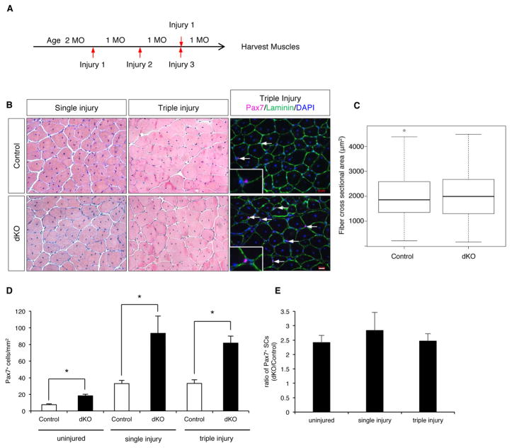
Figure 3. dKO Mice are Capable of Repetitive Muscle Regeneration.
(A) Protocol for assessing injury-induced regeneration.
(B) H&E-stained sections from control and dKO TA muscles subjected to one or three injuries; IF of Pax7 and laminin in TA sections. Arrows indicate SCs. Insets show higher magnification. Scale bar, 20μm.
(C) Box and whisker plots for cross-sectional areas of fibers from the regenerated region of TA after triple injury.
(D) Quantification of Pax7+ cells in TA sections from control and dKO mice prior to injury and after single and triple injury. *, p<0.005, means±SD.
(E) Ratio of Pax7+ cells in dKO vs. control mice prior to injury and after single and triple injury.

