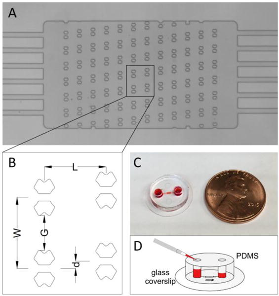
Figure 1.

A microfluidic device for arraying single cells. (A) The microarray of the butterfly-shaped traps. Each trap has a central aperture of 10 μm wide. The scale bar is 200 μm. (B) The detailed geometry of the trapping array. (C) The cell trapping microfluidic device is bonded to a glass coverslip (15 mm in diameter). (D) The hydrodynamic pressure drives the fluid flow through the device, which is governed by the height difference between the two fluid columns.
