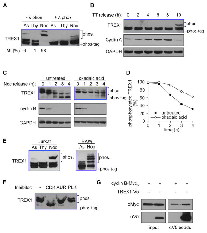
Figure 1. TREX1 Is Phosphorylated during Mitosis.
(A) HeLa cells were grown asynchronously (As), arrested with thymidine in interphase (Thy), or arrested in mitosis with nocodazole (Noc). Mitotic index (MI) measured by counting Phosphorylation of histone H3 serine 10 (p-H3S10) stained cells by fluorescent microscopy is indicated on the bottom. TREX1 phosphorylation was assayed using immunoblot of Phos-Tag PAGE (same throughout).
(B) HeLa cells were arrested in G1/S transition with double thymidine treatment (TT). Cells were released and collected at indicated time points on top. Mitotic entry is indicated by degradation of cyclin A. Loading control, GAPDH.
(C and D) HeLa cells were arrested in mitosis by thymidine/nocodazole treatment followed by okadaic acid (PP1/2 inhibitor) treatment, or not. Cells were released by washing out nocodazole and collected at the indicated time points. Mitotic exit is indicated by degradation of cyclin B. Loading control, GAPDH. Representative immunoblots shown in (C). Densitometry analysis of hyper-phosphorylated form of TREX1 is shown in (D).
(E) Jurkat T cells or RAW267.4 mouse macrophages are treated as in (A) to enrich cells arrested at interface or mitosis. TREX1 phosphorylation is detected by immunoblot of Phos-Tag PAGE.
(F) HeLa cells were arrested in mitosis by thymidine and nocodazole. Cells were treated 1 hr with RO3306 (CDK), ZM447439 (AUR), and BI2536 (PLK).
(G) TREX1-V5 and cyclin B-Myc6 were overexpressed in 293T cells arrested in mitosis by no-codazole. Cell extracts were immunoprecipitated using anti-V5 agarose beads. Cyclin B co-immunoprecipitation was detected using anti-Myc antibody. Data are representative of at least three independent experiments.

