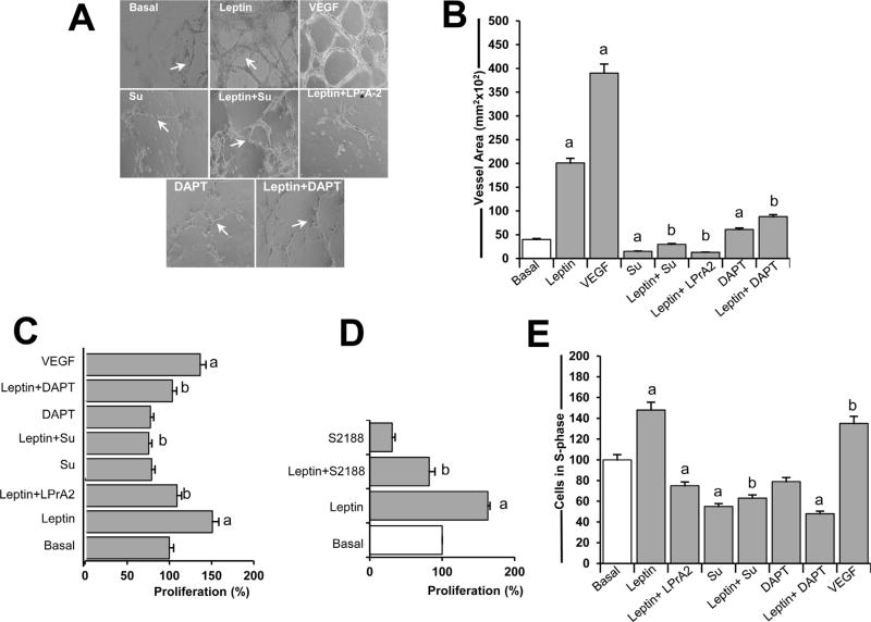
Fig 1. VEGFR-2 and Notch activities are essential for leptin-induced cell proliferation, s-phase progression, and tube formation in endothelial cells.
A. Leptin-induced tube-like formation in endothelial cells. Tube-like formation was analyzed in human umbilical vein endothelial cells (HUVEC) incubated in medium without (Basal) or containing leptin, VEGF, and inhibitors of VEGFR-2, leptin, and Notch. B. Quantitative determination of leptin-induced HUVEC tube-like formation. The graph shows tube-like formation calculated as vessel area (mm2) using Image Pro Plus software. The visible tubes were counted using the software, and the analyses of vessel average length and area were performed. HUVEC were cultured for 24 h in 96 well plates containing growth factor-reduced matrigel. Cells were treated with leptin (1.2 nM) and inhibitors of VEGFR-2 (SU5416; 5µmol/l), Notch (DAPT; 5 µmol/l), and leptin receptor (LPrA2, 20 nM), and positive control VEGF (25ng/ml).C. Leptin induces proliferation of endothelial cells. Results of MTT assay from HUVEC incubated as described in A. Absorbance was determined at 540 nm, and data was evaluated with Spectramax software. D. Inhibition of gamma-secretase blocks leptin-induced HUVEC proliferation. HUVEC were incubated with leptin (0 and 1.2 nM) and S2188 (10 nM; Notch inhibitor), and proliferation was determined via MTT assay. E. Leptin induces S-phase progression in HUVEC. HUVEC were cultured as described in 12-well cell culture plates for 24 h, and cell cycle progression was determined via Cellometer analysis (Nexelom). Quantitative analysis of propidium iodide-bound to DNA in S-phase gated cells was recorded. Data is presented as an average ±s.d. from three independent experiments. a: p<0.05 when compared to basal. b: p<0.05 when compared to endothelial cells treated with leptin. Su: SU5416. Magnification: 10×.

