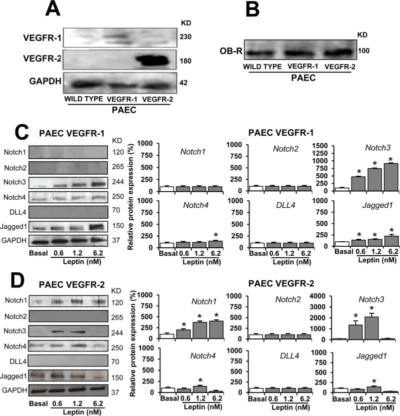
Fig. 4. VEGF receptors are involved in leptin-induced Notch expression in endothelial cells.
A. VEGFR expression in PAEC. WB representative results of VEGFR in PAEC transfected with VEGFR-1 and VEGFR-2. GAPDH was used as a loading control. B. Leptin receptor (OB-R) expression in PAEC. WB representative results from OB-R immunoprecipitation (IP) in PAEC wild type, and PAEC transfected with VEGFR-1 and VEGFR-2. Cells lysates (25 µg protein) were incubated at 4°C for 16 h with anti-OB-R antibody (0.5 µg).C.Leptin induction of Notch proteins in PAEC-VEGFR-1. Western blot (WB) representative results of leptin-induced Notch proteins in PAEC transfected with VEGFR-1. D. Leptin dose-dependent induction of Notch proteins in PAEC-VEGFR-2. PAEC transfected with VEGFR-1 and VEGFR-2 were cultured in medium containing leptin (0.6,1.2, and 6.2 nM) for 24 h. Cell lysates were used to determine Notch protein expression after treatment. GAPDH was used as a loading control. Histograms show densitometric analysis of protein expression normalized to GAPDH as determined using NIH image J software. Relative protein expression was calculated as percentage to basal. WB analysis did not show basal or leptin-induced expression of Notch proteins in wild type PAEC. Protein G-agarose beads were added for IP/WB analysis. Data is presented as an average ± s.d. from three independent experiments. * p<0.05 when compared to basal.

