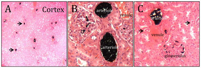
Figure 6.
Histology of embolized rabbit renal arteries. A) Low magnification cross-section through the cortex. The scattered black dots (arrows) are occluded arterioles. B) Higher magnification reveals occlusion of glomeruli capillaries (white arrow). C) The embolic coacervate was not present in venules or urine ducts (black arrows).

