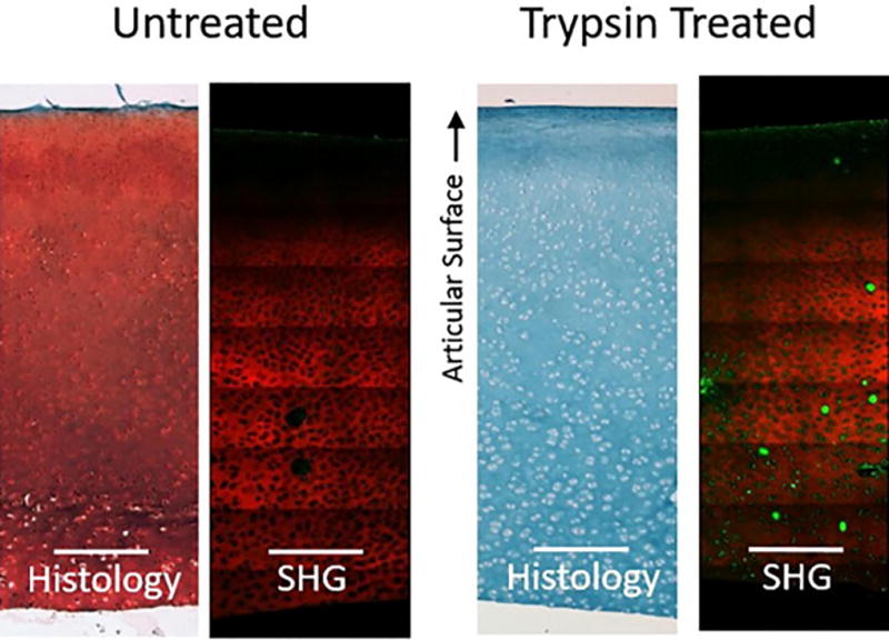
Figure 6.

Histology and SHG/TPF images of articular cartilage demonstrate GAG depletion due to trypsin treatment but little change to collagen matrix. Safranin-O/Fast Green histological stain on paraffin embedded samples. Fresh samples imaged with SHG for collagen type II artificially colored red and TPF artificially colored green.
