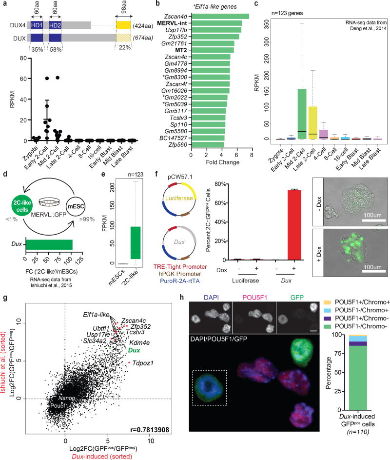
Figure 3. Mouse Dux, a functional ortholog of human DUX4, activates a ‘2C’ transcriptional program and converts mESCs to a ‘2C-like’ state.
(a) Sequence level comparison of DUX4 and DUX (top panel) and the normalized expression of Dux in pre-implantation mouse embryos (RNA-seq data from Deng et al., 2014) (bottom panel). (b) Bar graph displaying the top 15 differentially-expressed genes and repeat elements (bold) following ectopic Dux expression in mouse embryonic stem cells (mESCs) [two replicates per condition]. (c) Relative expression of Dux-induced genes (n=123) in the pre-implantation mouse embryo. (d) Diagram of mESC metastability (top panel) and the enrichment of Dux in ‘2C-like’ cells relative to conventional mESCs (bottom panel). (e) Expression of Dux-induced genes (n=123) in ‘2C-like’ cells compared to conventional mESCs. (f) Diagram of doxycycline-inducible lentiviral constructs stably integrated into mESCs (left panel) and their effect (after 24hrs of dox administration) on MERVL::GFP reporter expression evaluated by flow cytometry (middle panel) and live imaging microscopy (right panel) [four biological replicates per condition. Error bars, s.d]. (g) Dot plot showing per gene differential expression in Dux-induced MERVL::GFPpos cells (over MERVL::GFPneg cells), x-axis; compared with per gene differential expression observed in spontaneously converting ‘2C-like’ cells, y-axis. (h) Immunofluorescence quantifying the loss of pluripotency (e.g. POU5F1 protein) and chromocenters in mESCs following ectopic Dux expression (n =110 cells). Scale bar, 10um.

