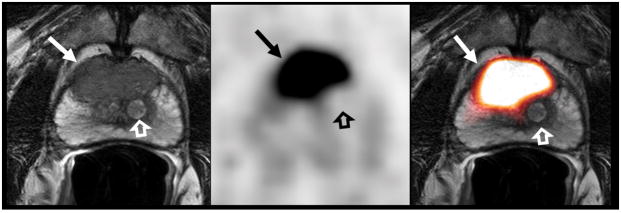
Figure 1.
73-year-old man with a serum PSA level of 38.6 ng/mL. T2W MR image (A), axial 18F-DCFBC PET image (B) and fused MRI/PET image (C) demonstrate a large low-signal-intensity focus in the anterior mid-central gland (arrow), which shows intense 18F-DCFBC uptake with SUVmax up to 16.3. Histopathology confirmed a Gleason score 4+5 prostate cancer. The BPH nodule (open arrow) and the normal prostate tissue does not demonstrate abnormal DCFBC uptake.

