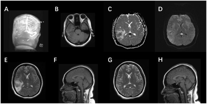
Figure 1.
(a–h): There is no flow within the right transverse sinus and sigmoid sinus of the source image of magnetic resonance venography (MRV) (a). On T1-weighted image (b) the thrombus within the right transverse sinus is directly visualized as hyperintense clot. Brain MRI showing an oval lesion in the SCC, hyperintense on DWI (c) and T2-weighted (e), and hypointense on the ADC map (d). Sagittal T1W1 (f) displays the same lesion, with slight hypointense signal. Axial T2W1 (e) shows mixed intensity signal in the right temporal lobe, indicating venous thrombosis with secondary hemorrhagic infarct. In the follow up 2 weeks later, note the regression of hemorrhagic lesion in T2W1 (g). The abnormal signal in corpus callosum also remitted completely (g, h).
ADC, apparent diffusion coefficient; DWI, diffusion-weighted imaging; MRI, magnetic resonance imaging.

