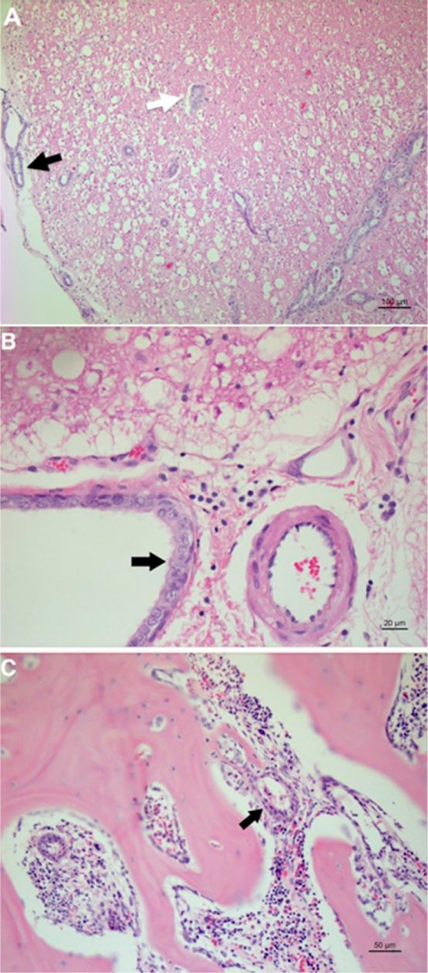
Figure 4.

Histopathological features on post-mortem examination, haematoxylin and eosin staining: (a) multifocal aggregates of neoplastic epithelial cells forming tubules infiltrate the leptomeninges (black arrow) and white matter of the spinal cord (white arrow); (b) detail of cilia within the apical border of neoplastic cells infiltrating the spinal cord white matter (black arrow); (c) multifocal aggregates of neoplastic cells within the bone marrow of vertebral bone (black arrow)
