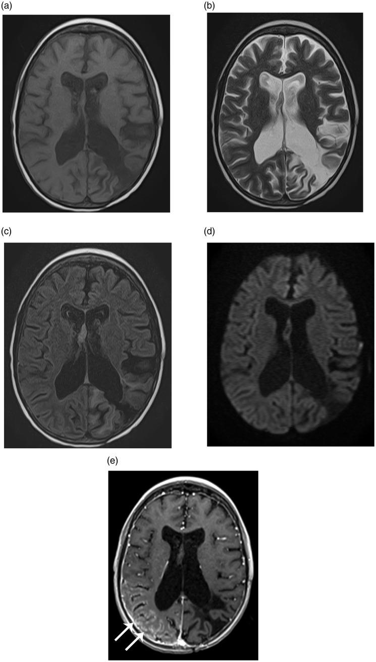
Figure 1.
Magnetic resonance (MR) images of the brain at the time of admission demonstrate gyriform enhancement (arrows in e) in the right parieto-occipital region without diffusion restriction. Note post-surgical encephalomalacia in the left parietooccipital region. (a) Axial T1-weighted image. (b) Axial T2-weighted image. (c) Axial fluid attenuation inversion recovery (FLAIR). (d) Axial diffusion-weighted image. (e) Axial T1-weighted post contrast image.

