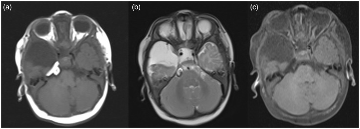
Figure 1.
A lesion within the right Meckel’s cave appearing hyperintense on T1-weighted imaging (WI) (a) and T2WI (b) showing suppression of signal intensity on T1 fat-suppressed sequences (c), suggesting diagnosis of Meckel’s cave lipoma. Also subcutaneous lipoma is seen over the right temporal region.

