Figure 1.
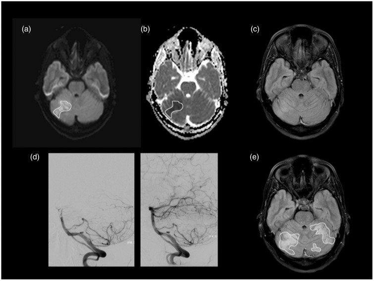
Patient 5, with occlusion at the distal basilar artery, was admitted with an NIHSS of 5. MRI scan was performed seven hours after symptom onset showing areas of restricted diffusion on right cerebellar hemisphere representing acute ischemia involving the superior cerebellar artery territory. We manually drew several ROIs including areas of hyperintensity at b1000 (a), subsequently imported to ADC map (b). The minADC value in this patient was 0.323 × 10⁻³mm2/s. A faint high signal is already seen on FLAIR at the corresponding location (c). Successful recanalization (TICI = 3) with mechanical thrombectomy using a stent retriever device was achieved three hours after initial MRI (d). The follow-up MRI was performed on day 4 and showed extension of the ischemic area to both cerebellar hemispheres superiorly (e). The patient had an NIHSS of 1 and mRS of 1 at discharge and no neurological deficits at three-month follow-up (mRS = 0). NIHSS: National Institutes of Health Stroke Scale; MRI: magnetic resonance imaging; ROI: region of interest; FLAIR: fluid-attenuated inversion recovery; ADC: apparent diffusion coefficient; TICI: Thrombolysis in Cerebral Infarction score.

