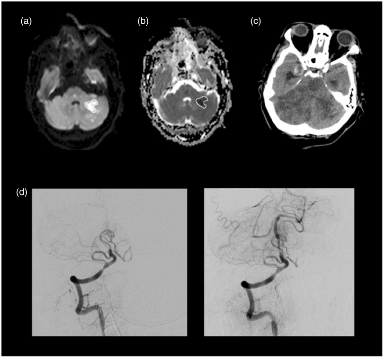
Figure 2.
Patient 11, with occlusion at the proximal basilar artery, was admitted with an NIHSS of 7. MRI scan showed multiple small areas of restricted diffusion representing acute ischemia on midbrain and pons bilaterally and on the left cerebellar hemisphere and temporo-occipital medial region. We did not take into account foci smaller than 1 cm when manually drawing ROIs including areas of hyperintensity at b1000 (a), subsequently imported to ADC map (b). MinADC measured in this patient was 0.219 × 10⁻³mm2/s. Despite successful recanalization (TICI = 3) with mechanical thrombectomy using a stent retriever device and balloon for dilatation (d), the follow-up CT two days after showed an extensive ischemic area involving both cerebellar hemispheres and midbrain (c), complicated with hemorrhagic transformation and supratentorial hydrocephalus, that culminated on patient death. NIHSS: National Institutes of Health Stroke Scale; MRI: magnetic resonance imaging; ROI: region of interest; FLAIR: fluid-attenuated inversion recovery; ADC: apparent diffusion coefficient; TICI: Thrombolysis in Cerebral Infarction score; CT: computed tomography.

