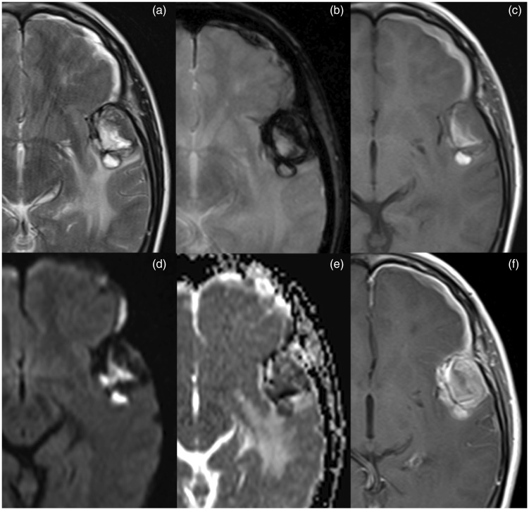
Figure 1.
Brain MRI. Lesion in the left temporal with heterogeneous signal in T2-weighted imaging (a) and enhancement (f); lesion has high signal components in T1-weighted imaging (c) compatible with haemorrhagic content and presents a peripheral rim of low signal in T2* (b) suggesting haemosiderin deposition. Diffusion-weighted imaging and apparent diffusion coefficient map (d and e) show heterogeneous signal due to the presence of blood products.

