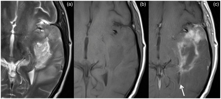
Figure 2.
Brain MRI. Primary gliosarcoma extending from the temporal cortical surface to the temporal horn of the lateral ventricle, heterogenous, but with predominant hyperintensity in T2-weighted imaging (a) and hypointensity in T1-weighted imaging (b); the enhancement is more ‘solid’ near the temporal cortical surface and more rim-like in the more central component; it reaches the vicinity of the ventricular system being possible to find ependymal lining enhancement (c, arrow).

