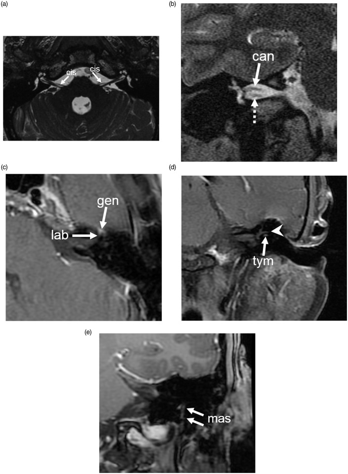
Figure 1.
Segments of facial nerve. (a) Axial FIESTA sequence shows the cisternal segment (cis) of the seventh cranial nerve emerging from the lateral brainstem between the pons and medulla at the level of the facial colliculus. (b) Coronal T2-weighted image shows the canalicular segment of the facial nerve (can) in the superior aspect of the internal auditory canal, with the cochlear nerve (dotted arrow) inferiorly located. (c) Axial gadobutrol contrast-enhanced SE T1 shows the labyrinthine (lab) and geniculate (gen) segments of the facial nerve. (d) Coronal gadobutrol contrast-enhanced SE T1 shows the tympanic segment (tym) of the facial nerve inferior to the lateral semicircular canal (arrowhead). (e) Coronal gadobutrol contrast-enhanced T1 (FSPGR) image shows the vertical mastoid segment (mas) of facial nerve extending from the posterior genu to the stylomastoid foramen. FIESTA: fast imaging employing steady-state acquisition; SE: spin-echo; FSPGR: fast spoiled gradient-echo.

