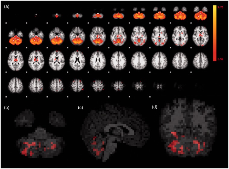
Figure 4.
Functional representation of cerebellar, with areas of thalamic, network. All first and second MRI scans were considered in the ICA (a). Corrected RS-fMRI maps in the axial (b), sagittal (c), and coronal (d) planes, comparing the second vs. the first MRI of the HIV-positive participants. Red voxels in (b), (c) and (d) represent areas with increased mean connectivity at the second MRI, encompassing the cerebellar vermis and hemispheres, in this network. MRI: magnetic resonance imaging; ICA: independent component analysis; RS-fMRI: resting-state functional magnetic resonance imaging; HIV: human immunodeficiency virus.

