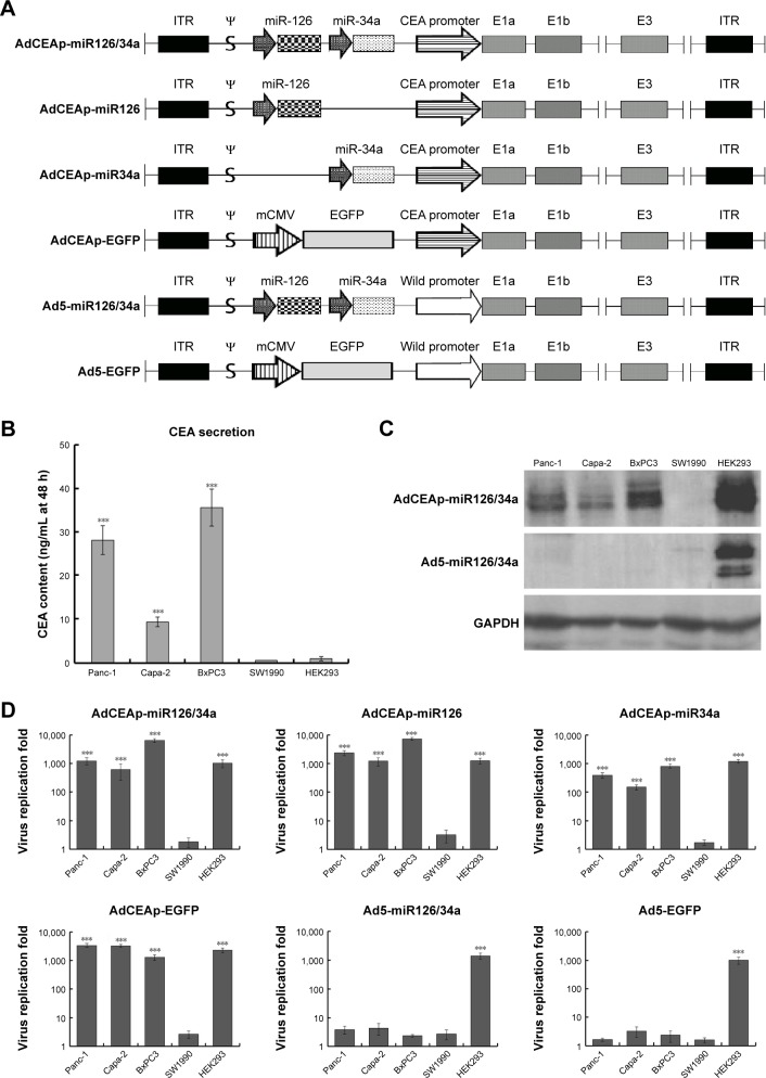
Figure 1.
Specific replication of the CEA promoter-regulated oncolytic adenovirus in PAC cells.
Notes: (A) Molecular structure diagrams of the constructed adenoviruses. ψ: adenovirus type 5 packaging signal. (B–D) Cell lines were planted in 6-well plates at 1×10⁶ per well and infected with the recombined adenoviruses at an MOI of 1 pfu/cell. (B) After 48 hours postinfection, cell culture supernatants were collected, and an electrochemiluminescence assay was used to detect CEA levels. ***P<0.001 versus the SW1990 group. (C) After 48 hours postinfection, cells were collected and used to detect E1a expression by Western blotting, with GAPDH as the loading control. (D) After 48 hours postinfection, cells were collected and the viral titers were quantified using the TCID50 assay. ***P<0.001 versus the SW1990 group.
Abbreviations: AdCEAp-miR126/34a, carcinoembryonic antigen promoter-driven oncolytic adenovirus; ITR, inverted terminal repeats; PAC, pancreatic adenocarcinoma; CEA, carcinoembryonic antigen; MOI, multiplicity of infection; TCID, tissue culture infectious dose 50; EGFP, enhanced green fluorescent protein.

