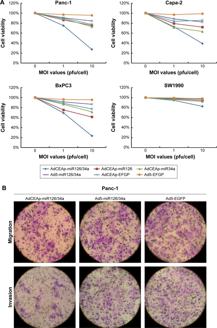
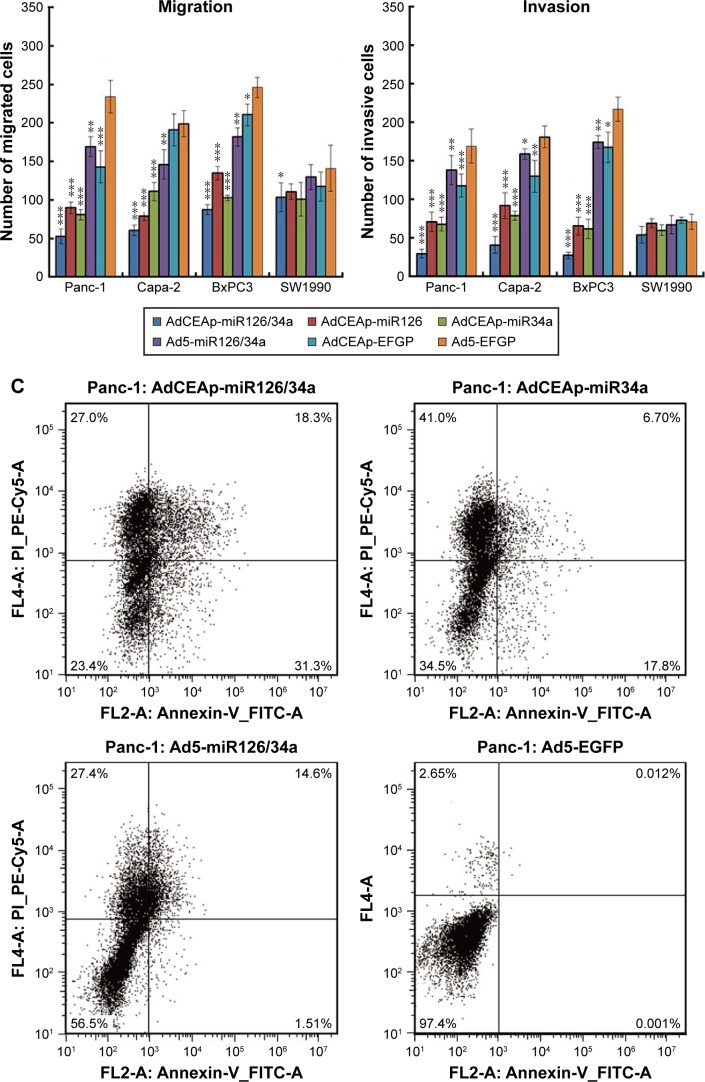
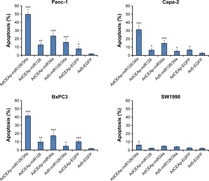
Figure 3.
Specific cytotoxicity of oncolytic adenovirus in PAC cells.
Notes: (A) Cells were seeded in 96-well plates at 1×10⁴ cells/well and infected with viruses at MOIs of 0, 1, and 10 pfu/cell. After 48 hours postinfection, cell viability was examined using the MTT assay. (B) Cells were planted in transwell chamber of 24-well plates at 5×10⁴ per well and treated with adenoviruses at an MOI of 1 pfu/cell. After 48 hours postinfection, the capacity of cell invasion and migration was assessed. The penetrated cells were counted within five high-power fields. Original magnification ×200; *P<0.05, **P<0.01, and ***P<0.001 versus the Ad5-EGFP-infected cells within the same cell line. (C) Cell lines were planted in 6-well plates at 5×10⁵ per well and infected with adenoviruses at an MOI of 1 pfu/cell. After 48 hours postinfection, cells were collected and stained with Annexin V/PI, and then analyzed by flow cytometry. The percentages of apoptotic cells included the early apoptotic cells (FITC-positive and PE-Cy5-negative) and the late apoptotic cells (FITC-positive and PE-Cy5-positive). *P<0.05, **P<0.01, and ***P<0.001 versus the Ad5-EGFP-infected cells within the same cell line.
Abbreviations: AdCEAp-miR126/34a, carcinoembryonic antigen promoter-driven oncolytic adenovirus; PAC, pancreatic adenocarcinoma; CEA, carcinoembryonic antigen; MOI, multiplicity of infection; EGFP, enhanced green fluorescent protein; PI, propidium iodide; FITC, fluorescein isothiocyanate; MTT, methyl thiazolyl tetrazolium.


