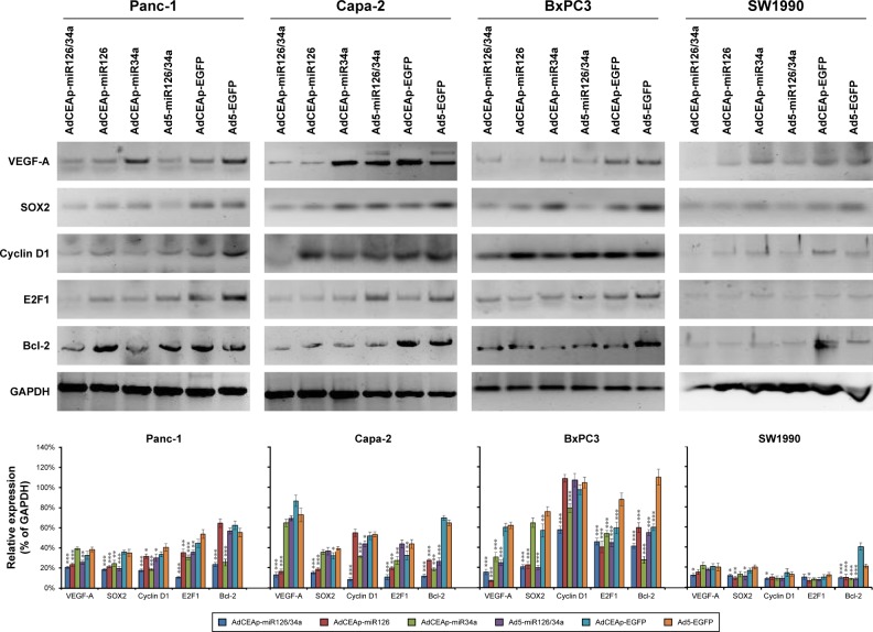
Figure 4.
Effect of miR-126 and miR-34a on their target genes.
Notes: Cells were planted in 24-well plates at 1×10⁶ cells/well and infected with adenoviruses at an MOI of 1 pfu/cell. After 48 hours, the harvested cells were examined for the expression of indicated proteins by Western blotting. The densitometry analysis of every band was normalized with GAPDH density. *P<0.05, **P<0.01, and ***P<0.001 versus the Ad5-EGFP-infected cells.
Abbreviations: AdCEAp-miR126/34a, carcinoembryonic antigen promoter-driven oncolytic adenovirus; CEA, carcinoembryonic antigen; MOI, multiplicity of infection; EGFP, enhanced green fluorescent protein; VEGF, vascular endothelial growth factor.

