Figure 5.
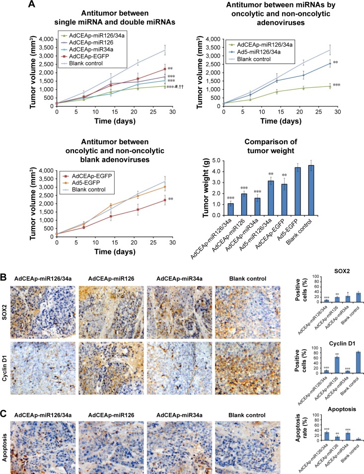
Antitumor efficacy of miR-126 and miR-34a expressed by oncolytic adenoviruses in PAC xenograft models.
Notes: (A) Nude mice were implanted with 1×10⁶ Panc-1 cells to establish xenograft models, and randomly divided into seven groups (AdCEAp-miR126/34a, AdCEAp-miR126, AdCEAp-miR34a, Ad5-miR126/34a, AdCEAp-EGFP, Ad5-EGFP, and blank control), n=5 for each group. The mice in each group were given intratumoral injections of the corresponding adenovirus at a total dose of 109 pfu. The blank control group was injected with the same volume of PBS synchronously. The tumor diameters were measured weekly and the tumor volumes were calculated. At day 28, the xenografted tumors were removed and weighed. **P<0.01 and ***P<0.001 compared with the blank control group, #P<0.05 compared with the AdCEAp-miR34a group, ††P<0.01 compared with the AdCEAp-miR126 group. (B) The paraffin-embedded sections of the xenografted tumors were prepared for examining the expression of SOX2 and cyclin D1 by immunohistochemistry. The percentages of positive cells were counted within 5 high-power fields. Magnification: 200×. *P<0.05, **P<0.01 and ***P<0.001 versus the blank control group. (C) Apoptosis was examined by the TUNEL assay. The percentages of positive cells were counted within five high-power fields. Magnification: 200×. **P<0.01, and ***P<0.001 versus the blank control group.
Abbreviations: AdCEAp-miR126/34a, carcinoembryonic antigen promoter-driven oncolytic adenovirus; PAC, pancreatic adenocarcinoma; CEA, carcinoembryonic antigen; EGFP, enhanced green fluorescent protein; PBS, phosphate-buffered saline; TUNEL, terminal-deoxynucleotidyl transferase-mediated dUTP nick end labeling; miRNA, microRNA.

