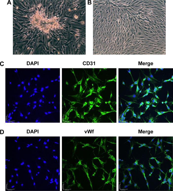
Figure 1.
Morphology and immunofluorescence in RCECs.
Notes: RCECs from rat retinal microvascular fragments were cultured in Endothelial Cell Medium. The RCECs fully released and migrated from the capillary fragments after culturing for 5 days (A). After 10 days, the cells showed a short fusiform or round morphology and formed contact-inhibited confluent monolayers with typical cobblestone morphology (B). The RCECs were positive for both CD31 (C, green fluorescence) and vWf (D, green fluorescence), as determined by an immunofluorescent assay. All nuclei were stained with DAPI (blue fluorescence). Magnification ×200.
Abbreviations: DAPI, 4′,6-diamidino-2-phenylindole; RCECs, retinal capillary endothelial cells; vWf, von Willebrand factor.

