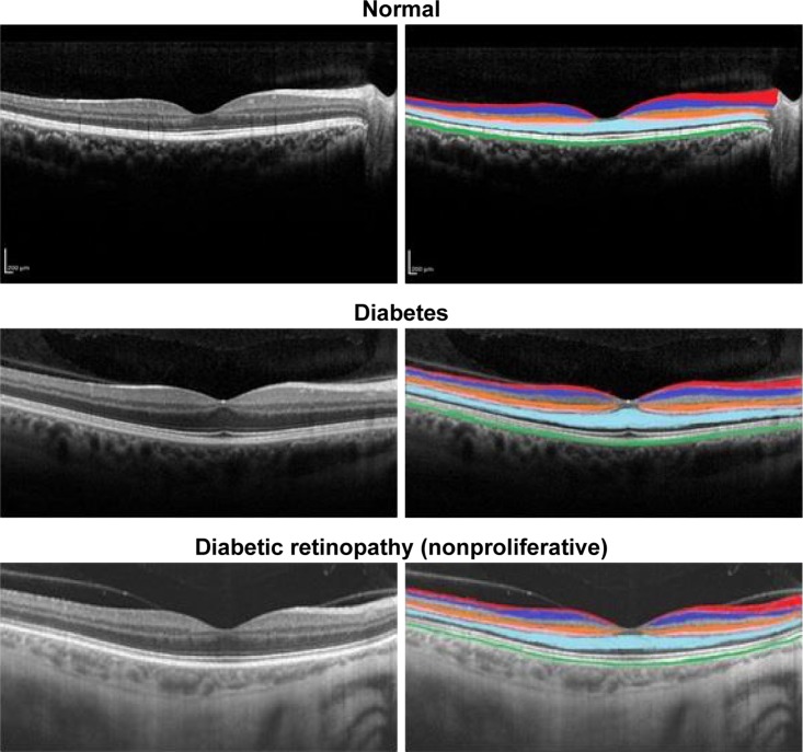
Figure 1.
Retinal OCT images showing six layers and their thicknesses highlighted in different colors.
Notes: RNFL, red; GCL, blue; INL, orange; OPL, pink; ONL + IS, sky blue; RPE, green. Scale bar 200 μm.
Abbreviations: GCL, ganglion cell layer; INL, inner nuclear layer; IS, inner segment layer; OCT, optical coherence tomography; ONL, outer nuclear layer; OPL, outer plexiform layer; RNFL, retinal nerve fiber layer; RPE, retinal pigment epithelium.

