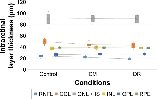
Figure 6.

Interconnected box plots showing intraretinal layer thickness measurements (in micrometers) in the pericentral area of the macula in patients with type 2 diabetes with no DR (DM) or DR compared to normal healthy individuals (NOM).
Note: The error bars indicate span of the data and also indicate the variability of data.
Abbreviations: DR, diabetic retinopathy; DM, diabetes mellitus; GCL, ganglion cell layer; INL, inner nuclear layer; IS, inner segment layer; NOM, normal healthy condition; ONL, outer nuclear layer; OPL, outer plexiform layer; RNFL, retinal nerve fiber layer; RPE, retinal pigment epithelium.
